Fluorescence imaging technology, as a vital tool in biomedical research, plays a key role in fundamental life science research, preclinical medical studies, and drug development due to its minimal tissue damage, absence of harmful electromagnetic radiation, and low-cost imaging equipment. With the rapid advancement of nanotechnology, fluorescent nanomaterials have emerged as a research hotspot in this field, owing to their unique optical properties, revolutionizing biological imaging.
Types and Characteristics of Fluorescent Nanomaterials
Fluorescent nanomaterials refer to ultra-small materials with fluorescence properties and at least one dimension in the 1-100 nm range. Their diverse types and unique characteristics provide versatile options for biological imaging.
Organic Fluorescent Dye-Based Nanoparticles
Organic fluorescent dyes, such as fluorescein and rhodamine derivatives, are commonly used as fluorescent probes in biomedical research. Combining organic dye molecules with nanoparticles can be achieved in two ways: first, by encapsulating the dye molecules within organic polymers or inorganic nanoparticles; second, by adsorbing the dye molecules onto the nanoparticle surface through chemical or physical methods.
This approach enhances the stability of dye molecules in biological environments, preventing their diffusion in tissues. Additionally, surface modifications with proteins or biomolecules enable specific labeling and fluorescence imaging of cells and tumor tissues. For example, Qian Jun et al. developed PpIX@SiO₂ nanoparticles by encapsulating the organic fluorescent molecule PpIX in silica nanoparticles, which exhibited excellent dispersibility in water and were suitable for in vitro cell and in vivo imaging.
Aggregation-induced emission (AIE) materials, discovered in 2001, feature unique molecular structures that exhibit significantly stronger fluorescence in the aggregated state than in dilute solutions, effectively addressing the fluorescence quenching issue of traditional organic dyes at high concentrations. For instance, our research group prepared stable nanomicelles (StCN@PEG) by encapsulating hydrophobic AIE materials (StCN) with amphiphilic polymers (mPEG-DSPE), which accumulated in tumor sites for in vivo fluorescence imaging.
Semiconductor Quantum Dots
Semiconductor quantum dots are typically composed of group II-VI (e.g., CdS, CdSe, CdTe) or group III-V (e.g., InP, InAs) elements, with particle sizes ranging from 1-10 nm. Due to their size being smaller than or comparable to the exciton Bohr radius, the internal electrons and holes are quantum-confined, transforming the continuous energy band structure into discrete energy levels, resulting in fluorescence upon excitation.
The fluorescence emission wavelength of quantum dots can be adjusted by size. Selecting appropriate materials and sizes allows for a wide range of fluorescence emission spectra. Compared to traditional organic fluorescent dyes, quantum dots offer broad absorption spectra, narrow emission spectra, resistance to photobleaching, and ease of surface modification.
In terms of synthesis, high-temperature pyrolysis is a classic chemical method. Professor Tang Fangqiong and her team at the Chinese Academy of Sciences synthesized CdSe quantum dots in a non-toxic oleic acid-liquid paraffin system, pioneering a “green” approach for group II-VI quantum dots. However, most semiconductor quantum dots contain heavy metals, raising concerns about potential biotoxicity and environmental pollution. Developing environmentally friendly quantum dots is a critical direction.
Carbon-Based Fluorescent Nanomaterials
Carbon-based fluorescent nanomaterials include nanodiamonds, carbon nanotubes, graphene oxide nanoparticles, and carbon-based quantum dots. Nanodiamonds can acquire excellent fluorescence properties through doping with elements like nitrogen or boron, but their preparation and doping challenges limit their applications.
The near-infrared fluorescence properties of carbon nanotubes make them widely applicable in in vivo animal imaging. For example, Dai et al. at Stanford University injected water-soluble carbon nanotubes into the bloodstream of live mice and obtained vascular imaging information using second near-infrared fluorescence imaging. However, the large size of carbon nanotubes makes them difficult to metabolize and raises potential toxicity concerns.
Graphene oxide (GO) nanomaterials exhibit good biocompatibility and ease of surface functionalization. Due to surface defects and oxygen-containing groups creating bandgaps, GO can produce fluorescence, making it suitable for in vitro cell and in vivo imaging.
Carbon-based quantum dots (CQDs) feature broad and continuous excitation spectra, high fluorescence stability, and tunable emission wavelengths. Composed mainly of C, H, O, and N, they exhibit excellent biocompatibility. Their synthesis methods include “top-down” and “bottom-up” approaches, showing broad prospects in biological fluorescence imaging.
Rare-Earth-Doped Upconversion Luminescent Nanomaterials
Upconversion luminescent nanomaterials are nanocrystals doped with rare-earth elements that can absorb two or more infrared photons and emit one visible or near-infrared photon. Compared to traditional luminescent materials, they offer lower cytotoxicity, higher biocompatibility, nearly zero background noise, long fluorescence lifetimes, and resistance to photobleaching.
Upconversion nanomaterials are widely used in biophotonics research. For example, Professor Prasad’s team at the University at Buffalo injected upconversion fluorescent nanomaterials into mice via the tail vein, achieving high-contrast deep optical imaging. Additionally, extensive research has been conducted on surface modifications, in vivo distribution studies, and biotoxicity analysis, paving the way for deep optical biotherapy.
Applications of Fluorescent Nanomaterials in Biomedical Imaging
Fluorescent nanomaterials are extensively applied in biomedical imaging, spanning from cellular to in vivo imaging, providing powerful tools for disease diagnosis, treatment, and research.
Development of Nanoprobes
Fluorescent nanoprobes exhibit unique optical properties due to quantum size effects and small-size effects. Compared to traditional organic fluorescent dyes, they offer superior excitation and emission characteristics, controllable optical bands through size and composition adjustments, high fluorescence intensity, stability, long lifetimes, and excellent biocompatibility. These advantages make them ideal for labeling lesions and targets and studying metabolic distribution in organisms.
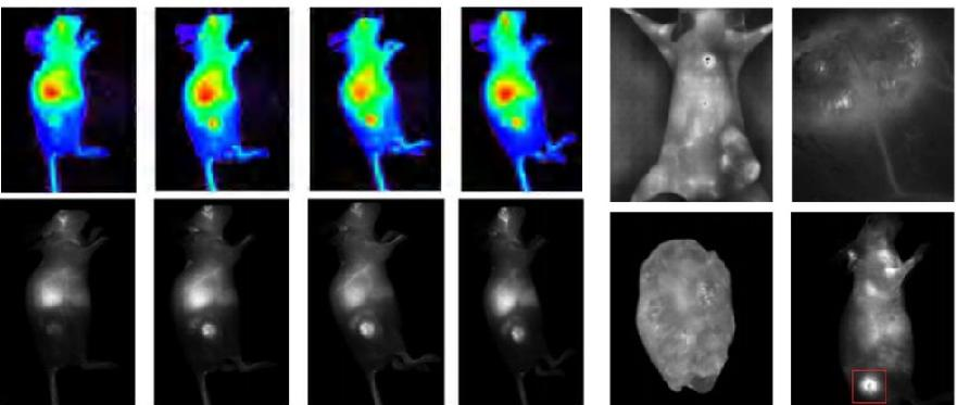
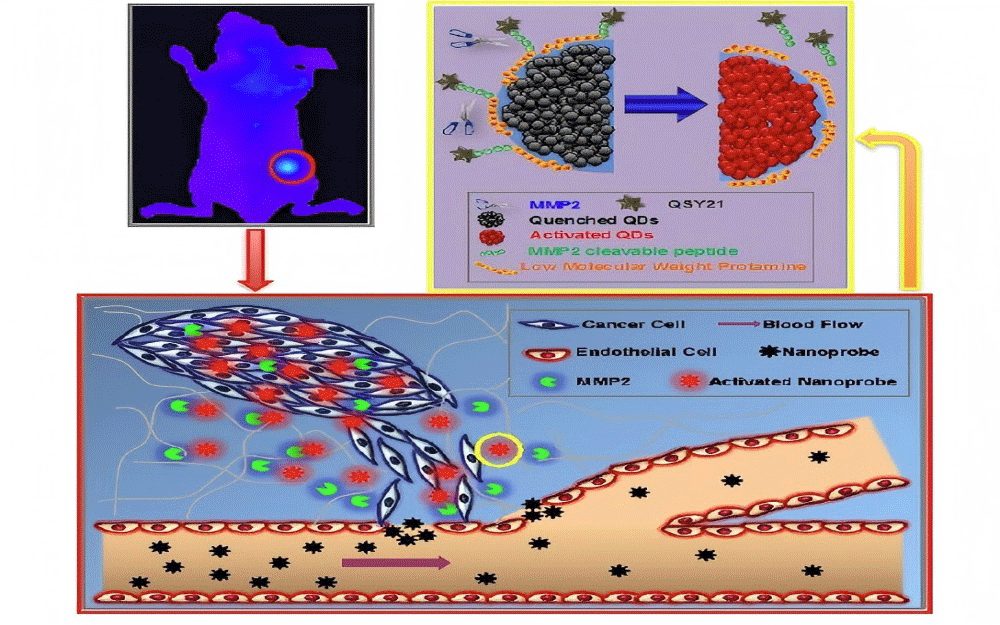
Probes developed for small-animal in vivo imaging include quantum dots, metal nanoparticles, upconversion nanomaterials, and semiconductor polymer nanomaterials. For example, Yaping Wang et al. developed an MMP-2-sensitive quantum dot nanoprobe to detect MMP-2-overexpressing tumors(Figure 1). Liyuan Pan reported an upconversion nanoprobe for targeted labeling of gastric tumors in vivo. Shuhendler et al. developed a composite nanoprobe to simultaneously monitor reactive oxygen species (ROS) and reactive nitrogen species (RNS) in mouse livers, evaluating drug-induced acute hepatotoxicity.
Research on Nanodrug Carriers
Nanodrug carriers have become a significant area in pharmaceutical research, offering advantages over traditional drugs: improved targeting through specific compound conjugation; enhanced membrane permeability and intracellular drug concentration via endocytosis; reduced drug concentration in non-target areas, minimizing side effects; and nanoscale sustained-release properties and high dispersibility, improving drug efficacy and utilization.
In vivo imaging of small animals is used to study the distribution and targeting of nanodrug carriers, evaluating efficacy and toxicity. Fluorescent or bioluminescent labeling of diseased cells can assess drug efficacy enhancement, while direct labeling of nanocarriers can track their distribution and localization in animals.
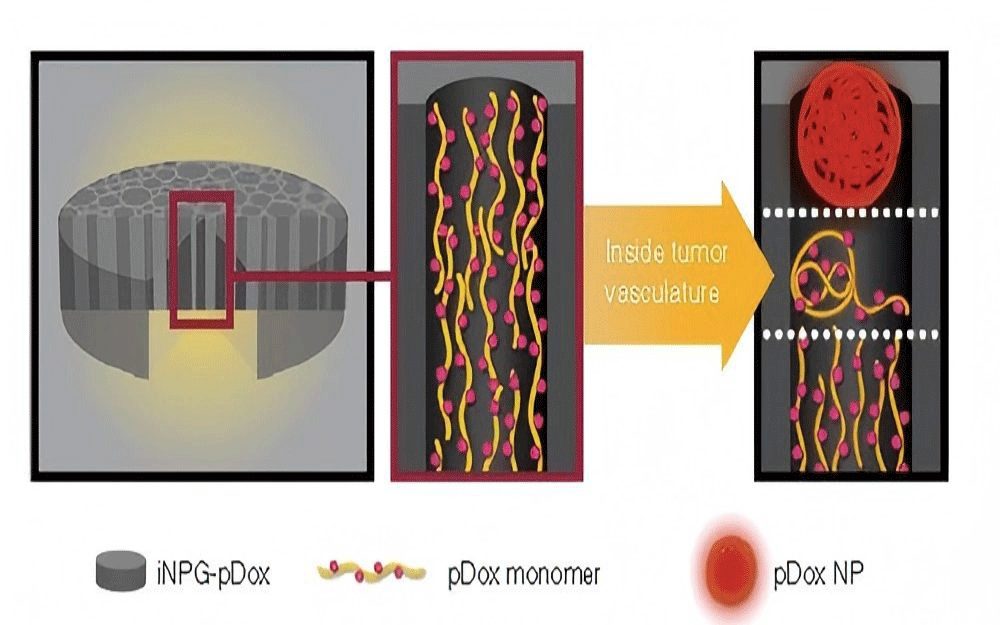
For example, Rong Xu et al. developed an injectable nanoparticle generator (iNPG-pDox) that overcomes biological barriers to efficiently target lung metastases of breast cancer, showing significant therapeutic effects(Figure 2). Hai Wang et al. designed a biomimetic nuclear cell nanodrug carrier (LC60S) co-loaded with ICG and DOX, achieving dual functions of targeted tumor therapy and real-time monitoring.
Biocompatibility and Safety Evaluation of Nanomaterials
The biocompatibility of nanomaterials is closely related to their entry into organisms. For nanoprobe development and nanodrug carrier construction, tissue compatibility is a critical consideration. All nanomaterials used in vivo require safety evaluations, with liver toxicity tests being a common method.
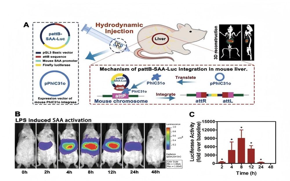
For instance, Zhang Y et al. at the Academy of Military Medical Sciences established a liver acute inflammation model to evaluate the hepatotoxicity of nanomaterials. Their study found that gold nanoparticles (GNPs) of different shapes and sizes variably activated SAA expression, with GNP50 inducing SAA activation via the NF-κB pathway(Figure 3).
Prospects and Challenges of Fluorescent Nanomaterials in Biomedical Imaging
Research on fluorescent nanomaterials for biomedical imaging is a multidisciplinary field, with new materials, technologies, and applications continuously emerging. Despite significant progress, challenges remain in translating these advancements into clinical practice.
Near-infrared fluorescence imaging represents a future trend in biological fluorescence imaging. Designing and preparing low-toxicity near-infrared fluorescent nanomaterials is a key direction for developing biomedical fluorescent probes. Multi-photon fluorescence imaging, requiring longer excitation wavelengths, reduces absorption and scattering in biological tissues, significantly improving resolution and imaging depth. Studies on the multi-photon fluorescence properties of nanomaterials are valuable for developing novel multi-photon-excited fluorescent probes.
Furthermore, comprehensive evaluations of cytotoxicity and organ toxicity are essential for nanomaterials. Research on material toxicity can guide the design and preparation of nanomaterials with better biocompatibility, offering new solutions for clinical applications such as tumor therapy.
As nanotechnology and biomedical research continue to converge, fluorescent nanomaterials will play an increasingly important role in biomedical imaging, driving breakthroughs in life sciences and medical development.