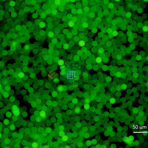
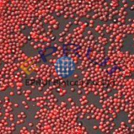
Fluorescent Polystyrene Microspheres
EPRUI supplies fluorescent polystyrene microspheres with high fluorescence intensity, stable performance and narrow particle size distribution.
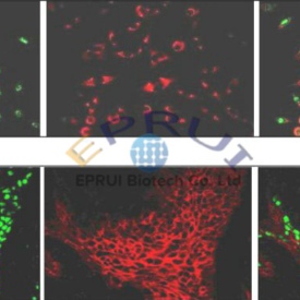
Our fluorescent polystyrene microspheres can be used for a wide range of applications including medical diagnosis, blood flow determination, tracing, in vivo imaging, calibration of imaging and flow cytometry instruments.
Our dyes are incorporated throughout the bead and not just on the surface, so they are relatively immune to photobleaching and other environmental factors. Dyes leak will not happen even after high-speed centrifugation. Carboxylated microbeads have a high density of homogeneous distribution of carboxylic acids on the surface, which makes carbodiimide (EDAC) and other water-soluble imines reagents covalently coupled protein and other amino-containing biomolecules easy to pass.
EPRUI Biotech Trusted By


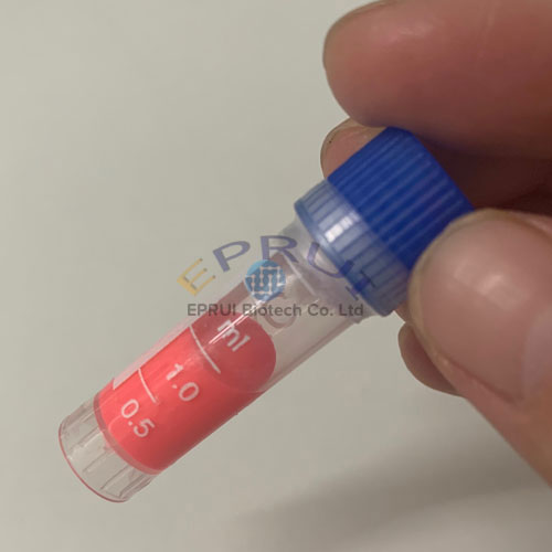
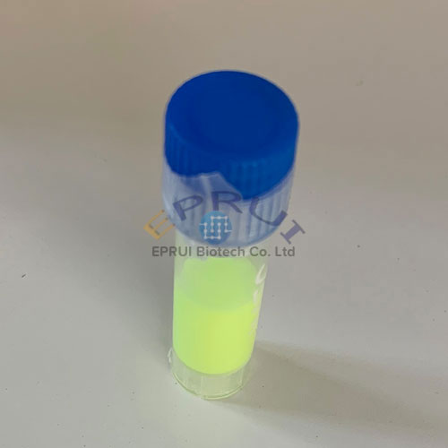
Fluorescent Microspheres Products
Buying Guide of Fluorescent Microspheres
1. What are Fluorescent Microspheres?
Fluorescent microspheres generally refer to the surface of the microspheres labeled with fluorescent substance (including surface coating) or the microspheres containing fluorescent substances (such as embedding or polymerization)
Fluorescent microspheres can be excited by external energy to excite fluorescence. The morphology of fluorescent microspheres can be any and spherical fluorescent microbeads are the most common type. In recent years, with the deepening of research on fluorescent probe technology, scientists are able to prepare a wide variety of particle sizes from nanometers to submicron fluorescent microspheres. Fluorescent microspheres have relatively stable morphological structure and luminescence behavior, and are less subject to external conditions such as solvent, heat, electricity and magnetism than pure fluorescent compounds.
The carrier of the fluorescent microsphere is mostly organic or inorganic polymer material. The fluorescent microspheres are divided into three categories based on the carrier as well as fluorescent substance.
- Inorganic/organic fluorescent microspheres
- Inorganic/inorganic fluorescent microspheres
- Organic/organic fluorescent microspheres.
Inorganic/organic fluorescent microspheres are further divided into:
- Inorganic materials as carriers and fluorescent substances are organic compounds
- Organic materials as carriers and fluorescent substances are inorganic compounds.
In addition, according to the position of the fluorescent substance in the carrier microsphere, the fluorescent microspheres can be divided into two categories.
- Fluorescent substance is on the surface of the carrier microspheres
- Fluorescent substance is inside the carrier microspheres
2. Fluorescent Microspheres Application
Due to the concentration of organic or inorganic substances capable of emitting fluorescence in a single microsphere, fluorescent microspheres have a wide range of applications in many fields such as biochemistry, biomedicine, clinical medicine, genetic analysis and optical instruments.
1) Application in marking and tracing
Fluorescent microspheres prepared by introducing one or more fluorescent substances on or inside polymer microspheres were first used to calibrate flow cytometry and as fluorescent quasi-microspheres for fluorescence microscope.
Gradually fluorescent microbeads are applied to cell labeling, biomolecule labeling, and tracking under active conditions.
Fix protein molecules, etc., and track their functionalization.
2) Application in detection
Mix the fluorescent microspheres with ligands on the surface that can bind to the test object and the radiolabeled test objects.
For a combined test object, its radiation can excite a fluorescent substance to generate fluorescence, while a test object that cannot be combined with microspheres emits radiation which will be submerged in water and cannot excite fluorescence due to its long distance. Fluorescent microspheres can carry many fluorescent molecules. Therefore, a weak stimulus can trigger a strong signal, so only a small amount of low-energy radiation can be used to generate a fluorescent signal avoiding radiation danger caused by the use of traditional radioactive microspheres and reduces the cost without losing the detection sensitivity
3) Application in immunoassay and drug screening
At present, fluorescent microspheres are most widely used in the field of biomedicine. The fluorescent microspheres that have been industrialized abroad are mainly used in this field.
It mainly includes high-throughput immunoassay, drug screening, and immobilized enzymes.
Fluorescent microspheres can be used as diagnostic reagents in immunoassays.
Fluorescent microspheres can be used as immobilized carrier for different detection objects in the detection of biological samples.
As a carrier, different fluorescent microspheres correspond to different capture antibodies and recognize different antigens,which realize the qualitative and quantitative detection of a variety of test antigens.
4) Application in immobilized enzyme and gene research
Enzymes can be fixed on fluorescent microspheres by physical adsorption or chemical bonding. Due to the three-dimensional shape of the enzyme is fixed, it not only has good pH stability, thermal stability and storage stability, and are easy to separate from the reactants, which can be reused and improve the use efficiency.
Therefore, the immobilization of enzymes or other biologically active substances on the fluorescent microsphere carrier is more conducive to its function.
5) Application in size standards
Fluorescent microspheres also have important applications in the measurement of cell flow cytometry and optical instruments, especially in the calibration of confocal microscopy.
In terms of cell flow cytometry, it is mainly used as a standard for measurement and for calibrating cell flow counters.
3. Preparation Methods of Fluorescent Microspheres
1) Physical adsorption method

The physical adsorption method is generally to dissolve a water-insoluble fluorescent substance in a water-soluble organic solvent such as acetone and ethanol, and then mix it with a water-dispersed system of a microsphere carrier. Is electrostatic) to the microsphere surface.
2) Self-assembly method

This method mainly uses inorganic or organic colloidal microspheres as a film-forming template. By electrostatic adsorption on its surface, inorganic or organic fluorescent nanoparticles can be obtained and assembled into membranes with different kinds of polyelectrolytes in an alternating manner. However, there are many steps to prepare fluorescent microspheres by this method, and the process is complicated.
Polyelectrolyte. This method is too complicated and is impossible for large-scale production. Later, People developed layer-by-layer self-assembly technology based on further research.
This method mainly uses inorganic or organic microspheres as the core.
Fluorescent microspheres are prepared by assembling fluorescent polyelectrolytes or fluorescent nanoparticles on their surfaces. The shell thickness of the obtained microsphere can be easily controlled by changing the number of cycles.
At the same time, the size and shape of the shell can be determined in advance by the size of the core used. This core-shell fluorescent microspheres have special properties with significant differences from nuclear (e.g. different chemical composition, good stability, high specific surface area, different magnetic and optical properties)
3) Embedding method

The basic principle of this method is to uniformly disperse the fluorescent material (including quantum dots) in the medium, using polymerization or microencapsulation methods
Preparation of fluorescent microspheres. If multiple fluorescent materials are uniformly dispersed in the same medium, the prepared fluorescent microspheres contain multiple fluorescent materials which can emit multiple fluorescent signals.
4) Chemical bonding

The fluorescent substance monomers or particles with active groups and the polymer microspheres with functional groups on the surface are chemically bonded to obtain”Large” fluorescent microspheres with a fluorescent substance on their surface.
5) Copolymerization

This method refers to fluorescent microspheres prepared by polymerizing fluorescent substance with a polymerizable functionalized organic monomer.
4. EPRUI Fluorescent Polystyrene Microspheres Features
- Dye molecules are incorporated throughout the bead making fluorescent signals more stable.
- At least 10% of dye concentration making sure high fluorescence intensity and brightness.
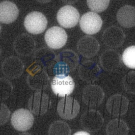
- Highly uniform particle size (CV<5%).Small batch difference and good repeatability.
- Customization of different functional groups and particle size (20nm-100um) is accepted.
5. Who are our customers?
Presently our customers are mainly from universities, research centers, labs as well as pharmaceutical companies, chromatographic column producers, diagnostic analysis and biotherapy related biotech companies.
Our university and research entity customers including:
- Harvard University
- Massachusetts Institute of Technology
- University of California – Berkeley
- Stanford University
- Vanderbilt University
- University of Minnesota
- The Rockefeller University
- Washington University
- OHIO University
- The University of Sheffield
- Imperial College London
- KAUST
- Newcastle University
- Physikalisch-Technische Bundesanstalt (PTB)
- University of Tübingen
- Fraunhofer IZFP
- ESPCI Paris
- Université de Tours
- IMT Atlantique
- ETH Zurich
- Fachhochschule Nordwestschweiz
- Korea University
- Seoul National University
- Nanyang Technological University
- National University of Singapore
- Singapore Institute of Manufacturing Technology
- Tsinghua University
- Fudan University
- Tongji University
Our company customers including:
- Illumina, Inc.
- Agilent Technologies
- BARD BRACHYTHERAPY INC
- LGC Biosearch Technologies Inc
- ChemGenes Corporation
- SamSung
- OSRAM
6. How to order
Please send e-mail to: sales@epruibiotech.com or call us by 86-21-64192663 for products inquires.
For detailed steps, please visit: How to Buy