Immunofluorescence (IF) is a biological technique that combines the ability of antibodies to specifically recognize antigens with the visualization properties of fluorescent dyes. It is used to detect the distribution, localization, and relative expression levels of specific proteins in tissues or cells. In simple terms, it uses “fluorescent-labeled antibodies” to locate “target proteins,” which are then observed under a fluorescence microscope. Due to its visualization capability, immunofluorescence is widely applied in life science research and clinical diagnostics.
However, conventional immunofluorescence assays have significant limitations: endogenous proteins and small molecule complexes in serum and biological fluids produce autofluorescence that overlaps with signals from conventional fluorophores, causing strong background interference and substantially reducing detection sensitivity. For example, biological samples such as serum and tissue fluid contain endogenous substances like tryptophan, tyrosine, NADH, and flavins. When excited by short-wavelength light (e.g., 300–400 nm), these substances emit fluorescence in the 400–600 nm range with a very short lifetime (nanosecond scale). Conventional fluorophores (e.g., FITC) also have very short fluorescence lifetimes, making it impossible for the detection window to avoid this interference.
To solve this problem, in 1983, scholars Soini and Kojola combined lanthanide element labeling with time-resolved fluorescence detection to create Time-Resolved Fluorescence Immunoassay (TRFIA). This technique uses lanthanide elements (most commonly europium Eu³⁺, terbium Tb³⁺, and samarium Sm³⁺), which have extremely long fluorescence lifetimes. It enables precise labeling of molecules such as proteins, enzymes, antigens, and antibodies, completely eliminating natural fluorescence interference from the temporal dimension, thus establishing a new generation of highly sensitive immunoassay platforms.
1. Detection Principle
Time-resolved fluorescence immunoassay relies on the unique fluorescence properties of lanthanides and immunochromatographic reactions to achieve quantitative detection. There are 15 lanthanide elements; the four ions commonly used in detection are samarium (Sm), europium (Eu), dysprosium (Dy), and terbium (Tb). Their fluorescence half-lives are as long as 10-1000us which is significantly longer than the 1-10ns background fluorescence of biological samples and completely eliminates natural fluorescence interference from the temporal dimension.
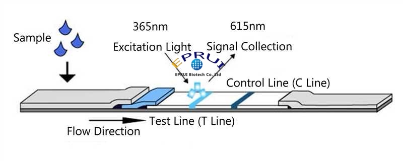
In rapid POCT detection, after the test sample is added to the sample application pad, the target antigen/antibody binds to the corresponding antibody/antigen labeled with fluorescent nanospheres. The complex moves along the chromatographic membrane by capillary action. When it reaches the test zone, a fluorescent microsphere-antibody-antigen-antibody sandwich complex is formed and immobilized on the test line (T line). Unbound labeled material continues to migrate to the control line (C line) and binds to the secondary antibody.
After the immunoreaction is complete, the detection zone is excited with 365 nm UV light. The lanthanide label emits high-intensity, long-lifetime fluorescence at approximately 615 nm. The instrument delays signal acquisition, allowing the short-lifetime background fluorescence from the sample to fully decay before reading only the long-lifetime specific fluorescence, achieving signal separation in the time dimension. Finally, the concentration of the analyte is calculated based on the fluorescence intensity ratio between the T line and the C line.
II. Advantages of Time Resolved or Rare-Earth Fluorescent Microspheres
- Extremely Long Fluorescence Lifetime
The fluorescence decay time of ions such as europium and samarium can reach tens to hundreds of thousands of nanoseconds, which is far longer than that of biological background fluorescence. By using a delayed detection window, non‑specific signals such as matrix autofluorescence and scattered light from containers can be completely eliminated, significantly improving the signal‑to‑noise ratio and lowering the detection limit.
- Large Stokes Shift
The Stokes shift is the wavelength difference between the excitation peak and the emission peak. Taking europium as an example, the excitation wavelength is 365 nm and the emission wavelength is 610 nm. This large shift allows the excitation light and emission light to be easily separated by optical filters, avoiding excitation light crosstalk and enhancing signal purity.

- Narrow and Specific Emission Spectrum
The emission peaks of lanthanide elements have a narrow full width at half maximum (FWHM), typically concentrated at 610 ± 5 nm, with sharp and well‑symmetrical peak shapes. Through wavelength selection, specific signals can be accurately distinguished from stray light, further reducing background noise and improving detection specificity.
III. Experimental comparison of sensitivity
A comparison of the sensitivity and signal performance of our company’s (Shanghai EPRUI Biotech Bio) 200 nm and 300 nm time-resolved fluorescent microspheres against a leading brand’s 200 nm time-resolved fluorescent microspheres in an immunochromatographic system yielded the following results:
- Sensitivity at low concentrations
At the lowest detected antigen concentration of 0.78125 mg/mL, the T-line signal of EPRUI Biotech 200 nm microspheres (618) was much higher than that of the leading brand’s 200 nm microspheres (135), and the corresponding T/C ratio (0.0294) was 2.3 times that of the leading brand (0.0126). EPRUI Biotech 300 nm microspheres performed even better, with a T-line signal of 741 and a T/C ratio of 0.0321, representing an approximately 2.5-fold improvement over the leading brand, demonstrating stronger low-concentration signal amplification and detection sensitivity.
- Linearity and dynamic range consistency
In the medium-to-high concentration range (1.5625–400 mg/mL), the T/C ratios of all three microspheres showed a stable gradient increase with rising antigen concentration, with good linear correlation. In the 25–400 mg/mL range, the T/C values of 200nm and 300nm time resolved fluorescent microspheres from EPRUI Biotech were essentially comparable to those of the leading brand. At some concentration points (e.g., at 100 mg/mL, 300 nm time resolved fluorescent microspheres from EPRUI Biotech has a T/C of 2.74 vs. the leading brand’s 2.49), the signal response of EPRUI Biotech microspheres is superior, indicating that their quantitative performance over a wide dynamic range is equivalent to or better than that of the leading brand.
- Background signal and specificity
In the blank control (antigen concentration 0 mg/mL), both 200nm and 300nm rare earth or time resolved fluorescent microsphere from EPRUI Biotech exhibited low T-line background signals (32/117), with T/C ratios of 0.0014 and 0.0049, respectively. Compared to the leading brand (0.0056), the EPRUI Biotech’s 200 nm microspheres has a much lower background, indicating less non-specific adsorption in the immunochromatographic system and a better signal-to-noise

EPRUI Biotech has been deeply engaged in the field of monodisperse microspheres for over a decade. Its monodisperse microspheres, including polystyrene microspheres, PMMA microspheres, silica microspheres, fluorescent microspheres, streptavidin microspheres, and magnetic microspheres, have a wide range of applications and are core enabling materials in fields such as biomedicine, LCD flat panel displays, analytical testing, in vitro diagnostics, and instrument calibration.
All rights reserved by EPRUI Biotech Co., Ltd. Reprinting is permitted with the original webpage link attached. Violators reproducing the content without authorization will be held legally liable.